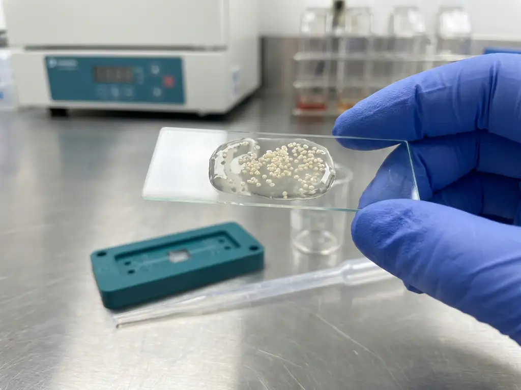
A preclinical probiotic adhesion assay tests how strongly and how long a strain binds to a gut mucus model under controlled conditions. Most workflows use a mucin binding test on coated plates, slides, or mucus-producing cell lines, then quantify retained cells after standardised washing. The key is choosing a relevant mucus source and validating the method so adhesion readouts support R&D decisions without being overinterpreted as clinical efficacy.
What does probiotic adhesion to gut mucus mean in preclinical testing?
In preclinical testing, probiotic adhesion to gut mucus means measuring a strain’s ability to attach to the mucus layer that coats the intestinal epithelium. This layer is rich in mucins, glycoproteins that create a hydrated barrier and a habitat for microbes. Adhesion typically reflects binding and short-term retention, sometimes used as a proxy for colonisation potential, but it is not the same as long-term colonisation or clinical benefit.
Common readouts include percent bound cells, cells per surface area, or apparent residence time in flow setups. Confounders to control include strain aggregation (false “high adhesion”), viability loss during handling, and variable expression of surface proteins due to growth phase or culture conditions.
How do you test probiotic adhesion to mucus in vitro?
Most in vitro mucus adhesion tests incubate a quantified probiotic suspension on a mucus-coated surface, wash away non-adherent cells, then quantify what remains. The simplest formats are microplate-based mucin binding tests, while more biorelevant options include flow-based adhesion or co-culture with mucus-producing cell lines such as HT29-MTX.
Typical workflow (plate or slide format)
- Coat wells or slides with mucin or extracted mucus, then block non-specific sites (for example with BSA).
- Prepare cells at a defined growth phase, standardise concentration, and select a quantification method (CFU, qPCR, or fluorescence labelling).
- Incubate under defined pH, ionic strength, bile exposure (if relevant), and time.
- Wash with a fixed protocol (volume, cycles, agitation), then quantify retained cells.
- Calculate adhesion as % retained versus input, or normalised to surface area.
Controls and replication
- Negative surface control (BSA-coated or uncoated wells) to estimate non-specific binding.
- Biological control strains (known low-adhesion comparator) and heat-killed cells to separate passive sticking from active surface interactions.
- Technical replicates and independent culture repeats to capture day-to-day variability.
Which mucus sources and models are most relevant for gut adhesion assays?
The most relevant mucus source depends on the intended site of action and target population. Purified porcine gastric mucin is widely used for convenience, but it may not match intestinal mucin composition or glycosylation. Intestinal mucins, human-derived mucus (biopsy-associated or other ethically sourced material), and synthetic hydrogels can improve fit-for-purpose modelling when you need tighter physiological alignment.
| Mucus source/model | Main strengths | Main limitations |
|---|---|---|
| Purified porcine mucin | Accessible, consistent, easy to standardise | May not reflect intestinal glycosylation and regional biology |
| Intestinal mucins/extracts | Closer to gut site chemistry | Batch variability, handling complexity |
| Human mucus (donor-derived) | Highest biological relevance | Limited supply, donor variability, ethics and logistics |
| Synthetic hydrogels | Tunable mechanics and chemistry | May miss key mucin motifs and native microstructure |
Also align conditions that strongly affect adhesion: pH, bile, ionic strength, shear, and mucin glycosylation patterns. For ileum versus colon questions, prioritise region-appropriate mucus and shear conditions.
What are the key pitfalls and how do you validate an adhesion assay?
The biggest risks in a mucin binding test are artefacts that look like adhesion. You validate by proving the assay has a usable dynamic range, is repeatable, and gives consistent results with orthogonal quantification. Set acceptance criteria up front so teams can compare strains, formulations, and process changes with confidence.
- Non-specific binding to plastic or blocking agents, solve with surface controls and optimised blocking.
- Aggregation inflating retention, monitor by microscopy or particle sizing and standardise dispersion.
- Plate effects (edge drying, coating gradients), mitigate with humidified incubation and randomisation.
- Over-washing or under-washing, lock a wash protocol and report it in full.
- Excipient interference (sweeteners, fibres, emulsifiers), test matrix-only controls.
- Viability loss, confirm input and post-assay viability where CFU is used.
Minimum reporting should include strain ID, growth phase, mucus concentration and source, incubation conditions, wash stringency, quantification method, and statistics.
How can ex vivo and dynamic gut models improve preclinical adhesion testing?
Ex vivo gut simulation and dynamic preclinical GI models improve adhesion testing by adding ecological realism: competition with resident microbiota, relevant metabolites, and time-resolved sampling under controlled anaerobic conditions. Rather than treating adhesion as a standalone KPI, these systems help connect retention behaviour to functional readouts without making clinical claims.
Practical ways to escalate beyond static assays include: testing adhesion after simulated digestion, assessing persistence in the presence of complex communities, and pairing adhesion with barrier-related markers in host–microbiome interaction set-ups. Escalate when you need to rank candidates across multiple donors, understand responder versus non-responder behaviour, or de-risk a formulation before committing to costly downstream work.
How Cryptobiotix helps with testing probiotic adhesion to gut mucus preclinically?
We help teams answer adhesion-related questions by combining GI simulation know-how with the SIFR® platform, so adhesion is interpreted alongside microbiome and functional context. Depending on your development stage, we can support:
- Study design that links a probiotic adhesion assay to downstream fermentation and mechanism questions across relevant cohorts, see our applications.
- Ex vivo experiments using SIFR technology to evaluate strain behaviour in complex communities, with controlled conditions and time-resolved sampling.
- Evidence packages aligned with decision-making needs, supported by our scientific evidence resources.
- Clear, actionable reporting that helps prioritise strains, doses, and formulations before larger investments.
If you want to discuss a fit-for-purpose gut mucus model and an efficient preclinical plan, contact us to align on objectives, controls, and readouts.
FAQ
Is high mucus adhesion enough to claim a probiotic will colonise the gut?
No. Adhesion indicates short-term binding under defined conditions. Long-term persistence depends on competition, nutrient availability, mucus turnover, and host factors, so adhesion should be treated as one input among several.
What is the best quantification method for an adhesion assay?
CFU counts measure viable adherent cells, qPCR measures total cells (live and dead unless viability dyes are used), and fluorescence is high-throughput but can be sensitive to labelling and quenching. Many teams use two methods to cross-check results.
How do you choose between mucin-coated plates and mucus-producing cell lines?
Mucin-coated plates are faster and easier to standardise for screening. Mucus-producing cell lines add biological context, including surface structure and host-derived components, but require tighter control of cell culture variability.